Imaging tests are an important part of the detection, diagnosis, and treatment of cancer. Imaging tests can be used for:
- Cancer screening to detect early signs of cancer when a person does not have any other signs or symptoms of cancer.
- Finding tumors that may be a cause of certain symptoms.
- Aid in determining whether a tumor is likely to be cancerous (malignant) or non-cancerous (benign)
- Determining if a cancer has spread to other body parts.
- Cancer treatment planning; to provide detailed images of exactly where the radiation therapy beams should be placed for treatment of a cancer.
- Surgical treatment planning, for tumors that need to be removed, Imaging tests are used to provide an exact location of the tumor prior to determining if it can be removed.
- Monitor the success of cancer treatment by creating images of a tumor to see if it has gotten smaller, stayed the same size, or has grown.
- Follow up once your cancer treatment has been completed. A follow up test can help to conclude if the cancer has been treated appropriately and is not reoccurring.
Many imaging tests require the use of a special dye, called contrast to highlight anatomy and provide additional information for diagnosis of diseases and tumors. Contrast for imaging tests is given in different ways, depending on the type of scan or x-ray test. You may be asked to drink contrast, it may be given by an enema through the rectum, or injected into an IV or port.
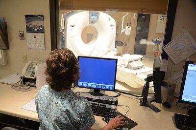
CT (computerized tomography) Scan

CT Guided Lung Biopsy – cross sectional CT image of needle placement into a lung nodule.
A CT scan is a test that uses x-rays and computers to created detailed images of the body. The images can be viewed as a slice, or cross section of the body and can also be constructed in the computer to produce a 3-dimensional picture. During a CT scan, you lie flat on a table which slides back and forth inside a donut shaped hole in the center of the scanner. CT scans are often the first test used to image tumors of the lungs, abdomen, liver, kidneys, pancreas, pelvis, spine, and brain. Additional tests are often recommended for further evaluation of a tumor found on CT.
In addition to providing an initial diagnosis of cancer, CT scans are used for very specific purposes throughout your cancer treatment; which may include:
CT Guided Biopsy is performed by a specialized doctor called an Interventional Radiologist. The CT machine is used to guide the Radiologist to take tissue samples of tumors. These tissue samples are then sent to a pathologist who will evaluate the cells to determine if the tumor is malignant or benign.
CT Simulation is a technique used to guide your Radiation Therapy Physician in planning treatment for cancer. During a CT simulation, you will lie on the CT table in the same position used for your radiation therapy treatments. Special positioning devices are often used for this scan. The detailed images produced by the CT scanner are put into a computer for your radiation therapy team to plan the precise location of where the radiation therapy beams will be placed.
MRI (Magnetic Resonance Imaging) Scan
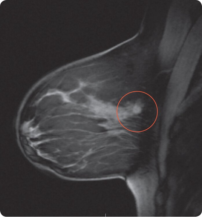
Breast MRI – cancerous tumor is shown with increased concentration of contrast.
MRI uses a strong magnetic field, radiofrequency signals, and sophisticated computer software to create cross sectional and 3-dimensional images of the body. MRI images provide excellent and clear detail of subtle soft tissue abnormalities. During an MRI scan, you lie on a table that is placed inside the scanner, which is shaped like a long cylinder. The MRI scanner will create loud noises while the magnetic field and radiofrequency signal are being activated. Earplugs and headphones are often provided to decrease the noise you will hear. Patients with pacemakers or certain medical device implants cannot have an MRI scan due to the strong magnetic field. The MRI technologist will carefully go over questions with you to ensure it is safe to perform the test.
MRI also has a very specific role in breast cancer screening and treatment strategies:
Breast MRI for Treatment Planning is frequently performed following an initial breast cancer diagnosis and prior to making any cancer treatment or surgery decisions. Breast MRI can provide specific information regarding the extent of a cancer and whether it has spread to other parts of the breast, or if the opposite breast is also affected.
Ultrasound Imaging
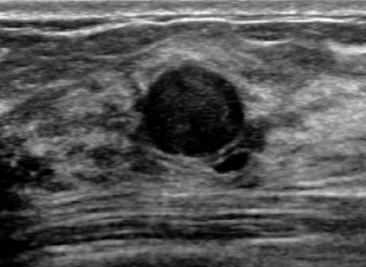
Breast ultrasound demonstrating a clear, fluid-filled, benign cyst.
An ultrasound creates images of organs in the body and is a very versatile test. The ultrasound machine sends out high frequency sound waves which are reflected off of the body structures as echoes of the sound waves. The echo signals are sent to a computer that creates an image from them. During an ultrasound exam, the technologist uses a hand-held probe, called a transducer, to obtain the images. The transducer is either moved over the body, or inserted into the body to produce the images.
Ultrasound can produce pictures of some diseases that are not well seen on exams that use x-ray. Using ultrasound, individual organs can be imaged to find tumors, or look for specific abnormalities of the tissue that can occur as a result of cancer. Ultrasound also uses a special technique called Doppler to listen to and see blood flow. This is an important part of cancer diagnosis, since tumors have blood flow that is different from normal tissue.
Ultrasound is frequently the first test ordered to diagnosis a wide variety of symptoms. And sometimes when an abnormal area is seen on another imaging exam, ultrasound is used to further evaluate the area or a biopsy is done with ultrasound to determine if the area is cancerous or not.
Breast Ultrasound is often the “next step” after an abnormal mammogram. Breast ultrasound helps to give detail of the area in question to determine if it is fluid filled (cystic) or solid (tumor), or needs additional testing for accurate diagnosis.
Ultrasound Guided Biopsy is performed to guide the Radiologist to take tissue samples of tumors. These tissue samples are then sent to a pathologist who will evaluate the cells to determine if the tumor is malignant or benign.
X-ray
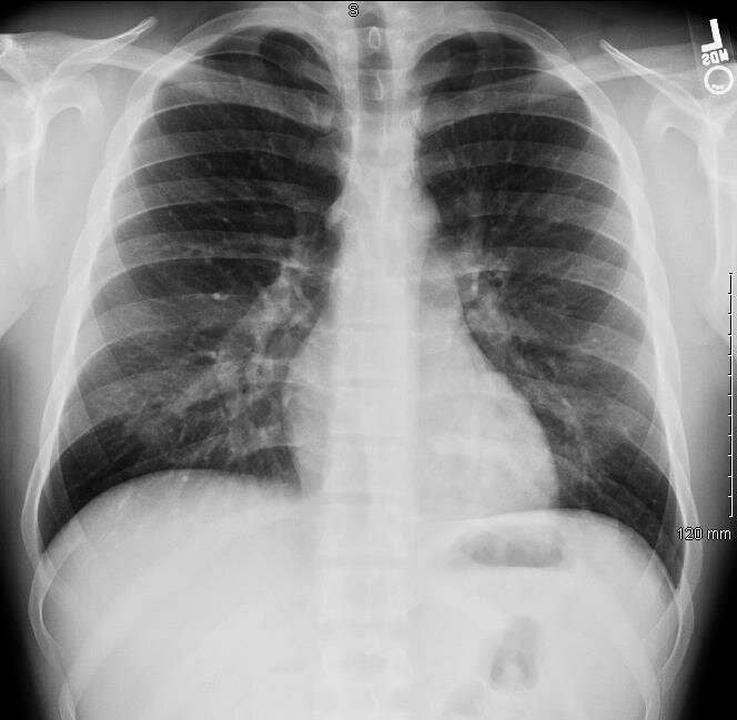
Chest x-ray
Many different diagnostic radiology exams use x-ray to diagnosis and evaluate disease. X-rays are fast and cost less than most other imaging tests, but do not provide the same detail that other tests can. A standard x-ray is sometimes used to initially evaluate symptoms or as a follow up to already known diseases.
X-ray tests may be used as part of your cancer treatment, or to help with the treatment other health symptoms that may occur during your cancer care.
Mammography
Mammography is an x-ray of the breast tissue, used to detect subtle abnormalities that may be a sign of cancer or to evaluate symptoms of breast disease. A mammogram is performed by taking x-ray pictures of the breast at different angles. The machine compresses the breast to spread the tissues apart so that the Radiologist can thoroughly evaluate all of the structures in the breast.
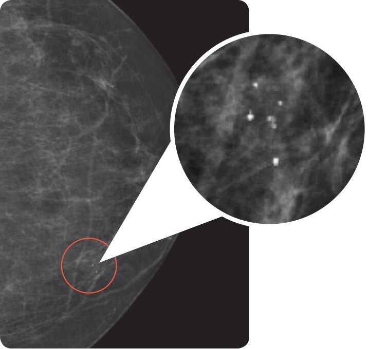
Mammogram – shows calcifications, an early sign of breast cancer
Mammography is used for the purpose of screening for breast cancer and diagnosing breast cancer.
Screening Mammography is recommended annually for women ages 40 and over, and earlier for women with certain risk factors for developing breast cancer. Screening mammography can detect cancer early, even before a lump can be felt. Early detection of breast cancer provides the most success for cancer treatment.
Diagnostic Mammography is used to evaluate abnormalities found on a screening mammogram, lumps that are felt by physical examination, changes in the breast tissue appearance, and other breast symptoms. A diagnostic mammogram takes additional pictures to provide images of the area in question.
Stereotactic Breast Biopsy is performed by a Radiologist who specializes in breast care. An advanced mammogram machine is used to locate an area in question and takes images to guide the Radiologist for removal of small tissue samples of the breast. These tissue samples are then sent to a pathologist who will evaluate the cells to determine if the area is malignant or benign.
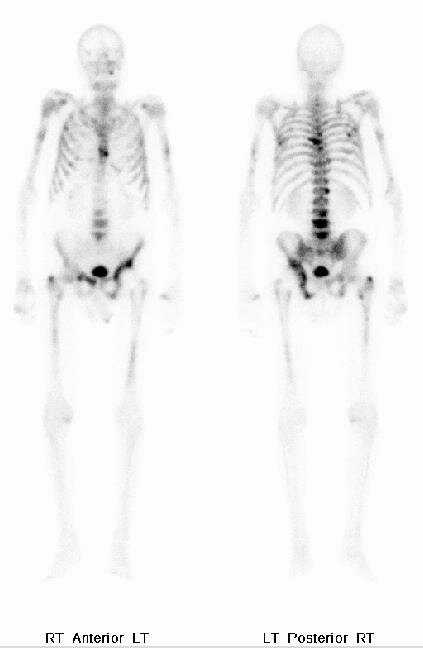
NM Bone Scan – “hot spots” are areas of abnormal uptake of the radioactive tracer.
Nuclear Medicine Scan
Nuclear Medicine scans create images based on the body’s chemistry and can provide information on how organs are functioning. During a Nuclear Medicine test, a small dose of a radioactive substance (sometimes also called a tracer) is injected in the body, usually by an IV. As the body tissue absorbs this radioactive material, a special camera picks up the pattern of radioactivity to create images. Depending on the test performed, several minutes to hours may be needed to allow the body adequate time to absorb the radioactive dose before images are taken.
PET (Positron Emission Tomography) / CT Scan

PET/CT scan showing a cancerous lung tumor.
A PET scan is type of Nuclear Medicine test which uses a special form of radioactive sugar. Cells in the body absorb this at different rates. Cancer cells grow quickly, and usually take in larger amounts of the radioactive sugar. Like Nuclear Medicine, the images will show “hot spots” in areas where the material has higher concentration. In a PET/CT scan, cross sectional images using a CT scanner are taken at the same time as the PET images. The combination of PET and CT provide very detailed information on the exact location and the cellular activity of a tumor.
PET/CT scans are often performed initially as a follow up to an abnormality found on another diagnostic imaging test. The initial PET/CT scan will diagnosis the likelihood that a tumor is cancerous and recommend further tests or biopsy if the area is at increased risk for being a cancer. If the area is determined to be less likely for cancer, then close monitoring with follow up imaging tests or other medical tests is sometimes necessary.
After cancer treatment has begun, a follow up PET/CT scan is sometimes performed. This will provide valuable detailed information on the tumor’s size and how well it is responding to treatment. A cancer treatment plan may be modified based on these results.
Interventional Radiology

X-ray showing placement of a central venous catheter in the Jugular vein for chemotherapy treatments.
Interventional Radiology is a specialized area of x-ray that has specific role in cancer diagnosis and treatment methods. A radiologist and team of technologist and nurses with particular training and skills perform a variety of procedures using either X-ray, Ultrasound, or CT to guide them during the procedure. Interventional Radiology can procedures provide both cancer diagnosis and treatment for complications
associated with associated with cancer. Some of these procedures include:
Biopsy is performed to remove tissue samples. The tissue samples are then evaluated for cancer cells.
Aspiration of fluid is done by inserting a fine needle into a collection of fluid that has gathered in the body. This can be done to relieve symptoms associated with increased fluid collection, or to evaluate the fluid for infection or cancer cells.
Tumor Ablation is a technique that uses special equipment to destroy tumors without removing them.
Central Venous Catheter placement, also called central lines, or ports, are used instead of IV’s for administering chemotherapy drugs. These devices can stay in the body for a long duration and are often recommended when frequent chemotherapy treatments or infusion are required, when multiple different drugs need to be provided at the same time, or when IV access is difficult to obtain.